
Cardiovascular questions are the leading commonly tested questions in pathophysiology exams especially if you are using books or such as Understanding Pathophysiology by Huether and McCance's. This is also evident in their respective test banks. Cardiovascular questions frequently cover heart failure, myocardial infarction, dysrhythmias, shock, and hypertension.
Let’s start from the basics, if you are asked in an exam to describe or define heart failure, most of the test banks have described the condition as the heart’s inability to meet the body’s demands, causing reduced cardiac output and fluid back-up in the lungs and veins.
Other related questions on the subject examine conditions such as systolic vs. diastolic failure, compensatory mechanisms, and signs like pulmonary edema. Acute and chronic valvular disorders, cardiogenic shock, and atherosclerosis (e.g. coronary artery disease) are also common, as is the underlying pathophysiology (e.g. plaque rupture, ischemia).
This is a classic topic that you will not miss in pathophysiology exams. Often, you will be examined on shock from any cause be it hypovolemic, cardiogenic, septic. Remember, in nursing shock is defined as life-threatening tissue hypoperfusion causing cellular dysfunction.
Get the Complete Huether & McCance’s Understanding Pathophysiology Test Bank
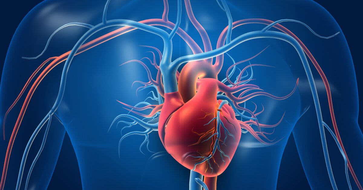
Commonly Tested Cardiovascular System Questions in Pathophysiology Exam
Sample questions and answers from Understanding Pathophysiology by Huether and McCance, 2nd Canadian Edition
Structure and Function of the Cardiovascular and Lymphatic Systems
1. The pericardium is:
a. the outer muscular layer of the heart.
b. the innermost layer of the heart chambers.
c. a membranous sac that encloses the heart.
d. the heart’s fibrous skeleton.
ANS: C
The pericardium is the membranous sac that surrounds the heart. The outer layer that acts as the fibrous skeleton of the heart is the myocardium. The innermost layer of the heart is the endocardium.
2. A function of the pericardium is to:
a. provide a barrier against extracardial infections.
b. improve blood flow through the heart.
c. play a role in cardiac conduction.
d. assist in cardiac contraction.
ANS: A
A function of the pericardium is to provide a barrier against extracardial infections. The pericardium does not improvNe UblRooSdIflNowGTthBro.uCghtMhe heart as it is on the outside. The inner portions of the heart control cardiac conduction. The muscular layers assist with cardiac contraction.
3. As a result of blockage in the pulmonary artery, blood would first back up into the: a. aorta.
b. left ventricle.
c. pulmonary veins.
d. right ventricle.
ANS: D
Blockage in the pulmonary artery would cause blood to back up into the right ventricle, not
the aorta since these two vessels do not communicate. The pulmonary artery and left ventricle do not communicate. Blockage in the pulmonary artery would not cause blood to back up into the pulmonary vein since the pulmonary vein takes blood to the left atrium.
REF: p. 571
4. Which chamber of the heart generates the highest pressure?
a. Right atrium
b. Left atrium
c. Left ventricle
d. Right ventricle
ANS: C
The left ventricle generates the highest pressure of all the heart’s chambers.
5. The internal lining of the cardiovascular system is formed by what tissue?
a. Tunica adventitia
b. Connective
c. Mesothelium
d. Endothelium
ANS: D
The endothelium, not the tunica adventitia, is the lining of blood vessels. Connective tissues help make up arterial walls but are not the lining of blood vessels. The mesothelium is a part of the pericardial cavity.
REF: p. 570
6. A 20-year-old underwent an echocardiogram to assess chest pain. Results revealed a congenital defect in the papillary muscles. Which of the following would the nurse expect to occur?
a. Closure of the semilunar valve
b. Backward expulsion of the atrioventricular valves
c. Closure of the atrioventricular valve
d. Backward expulsion of the semilunar valves
ANS: B
The papillary muscles are extensions of the myocardium that pull the cusps together and downward at the onset of ventricular contraction, thus preventing their backward expulsion into the atria. Defects in the papillary muscles would not affect either the semilunar or atrioventricular valves.
REF: p. 572
7. Which structures act as anchors for the atrioventricular valves?
a. Chordae tendineae
b. Great vessels
c. Coronary ostia
d. Trabeculae carneae
ANS: A
The atrioventricular valve openings are attached to the papillary muscles by the chordae tendineae. The great vessels are the vessels that bring blood to and out of the heart and are not attached to the chordae tendineae. The coronary ostia are openings in the aorta for the coronary arteries. The trabeculae carneae are a portion of the myocardium.
REF: p. 572
Alterations of Cardiovascular Function
1. A 75-year-old obese female presents to her primary care provider reporting edema in the lower extremities. Physical exam reveals that she has varicose veins. Upon performing the history, which of the following is a possible cause for the varicose veins?
a. Extreme exercise
b. Long periods of standing
c. Trauma to the deep veins
d. Ischemia
ANS: B
The probable cause of the patient’s varicose veins is gradual venous distention caused by the action of gravity on blood in the legs due to long periods of standing. Varicose veins are most likely due to long periods of standing leading to the action of gravity promoting venous distention. Exercise would help prevent this. Trauma can occur, but usually this affects the more superficial veins. Ischemia affects arteries not veins.
REF: p. 598
2. A 52-year-old male presents with pooling of blood in the veins of the lower extremities and edema. The diagnosis is chronic venous insufficiency, and an expected assessment finding of this disorder is:
a. deep vein thrombus formation.
b. skin hyperpigmentation.
c. gangrene.
d. edema above the knee.
ANS: B
Symptoms include edema of the lower extremities and hyperpigmentation of the skin of the feet and ankles but deep vein thrombi do not form. Edema in these areas may extend to the knees but not above. Gangrene does not occur in veins but in arteries.
REF: p. 598
3. Superior vena cava syndrome (SVCS), causing venous distention in the upper extremities, is a result of progressive superior vena cava:
a. inflammation.
b. occlusion.
c. distention.
d. sclerosis.
ANS: B
SVCS is a progressive occlusion of the SVC that leads to venous distention in the upper extremities and head. This distention is not a result of progressive inflammation, distention, or sclerosis.
REF: p. 599
4. A 50-year-old male with a 30-year history of smoking was diagnosed with bronchogenic cancer. He developed edema and venous distention in the upper extremities and face. Which of the following diagnosis will the nurse observe on the chart? a. Thromboembolism
b. Deep vein thrombosis
c. Superior vena cava syndrome (SVCS)
d. Chronic venous insufficiency
ANS: C
SVCS is a progressive occlusion of the superior vena cava that leads to venous distention in the upper extremities and head. Thromboembolism would not lead to the generalized symptoms described in the patient. Deep vein thrombosis would not lead to upper extremity symptoms. Chronic venous insufficiency would primarily affect one extremity.
REF: p. 599
5. When a patient is diagnosed with coronary artery disease, the nurse assesses for myocardial:
a. hypertrophy.
b. ischemia.
c. necrosis.
d. inflammation.
ANS: B
Coronary artery disease leads to myocardial ischemia. Coronary artery disease would not lead to hypertrophy, but not to ischemia, necrosis, or inflammation.
Alterations of Cardiovascular Function in Children
1. An 8-week-old infant presents to the pediatrician for a well-baby checkup. Physical exam reveals a murmur, and an echocardiogram confirms a ventricular septal defect. Which genetic disorder is likely to accompany this diagnosis?
a. Huntington disease
b. Color blindness
c. Down syndrome
d. Hemophilia
ANS: C
Down syndrome is the genetic factor that would most likely accompany the diagnosis of a congenital heart defect. Neither Huntington disease, color blindness, nor hemophilia is a
genetic disorder that would most likely accompany a diagnosis of congenital heart defect.
REF: p. 655
2. Intrauterine exposure to which factor could be responsible for a diagnosis of congenital heart disease?
a. Diabetes
b. Alcohol exposure
c. Viral infection
d. Dextroamphetamine
ANS: C
One of the identified causes of cardiac defects is an intrauterine viral infection, especially rubella. Diabetes contributes to large babies. Alcohol exposure contributes to abnormalities such as facial changes while exposure to dextroamphetamines leads to other abnormalities.
REF: p. 655
3. Coarctation of the aorta is the local narrowing of the aorta near the:
a. aortic valve.
b. ductus arteriosus.
c. diaphragm.
d. bifurcation into the common iliac arteries.
ANS: B
Coarctation of the aorta is narrowing of the aorta near the ductus arteriosus, not near the aortic valve, the diaphragm, or the bifurcation site.
REF: p. 656
4. A newborn is suspected of having coarctation of the aorta. Which of the following assessments would aid in diagnosis?
a. Cyanosis
b. Bounding pedal pulses
c. Cool arms
d. Weak or absent femoral pulses
ANS: D
0
691