Your cart is empty!
TEST BANK ILLUSTRATED DENTAL EMBRYOLOGY HISTOLOGY AND ANATOMY 4TH EDITION FEHRENBACH
Unit I: Orofacial Structures
1. Face and Neck Regions
2. Oral Cavity and Pharynx
3. Prenatal Development
4. Face and Neck Development
5. Orofacial Development
6. Tooth Development and Eruption
7. Cells
8. Basic Tissue
9. Oral Mucosa
10. Gingival and Dentogingival Junctional Tissues
11. Head and Neck Structures
12. Enamel
13. Dentin and Pulp
14. Periodontium: Cementum, Alveolar Bone, Periodontal Ligament
15. Overview of Dentitions
16. Permanent Anterior Teeth
17. Permanent Posterior Teeth
18. Primary Dentition
19. Temporomandibular Joint
20. Occlusion
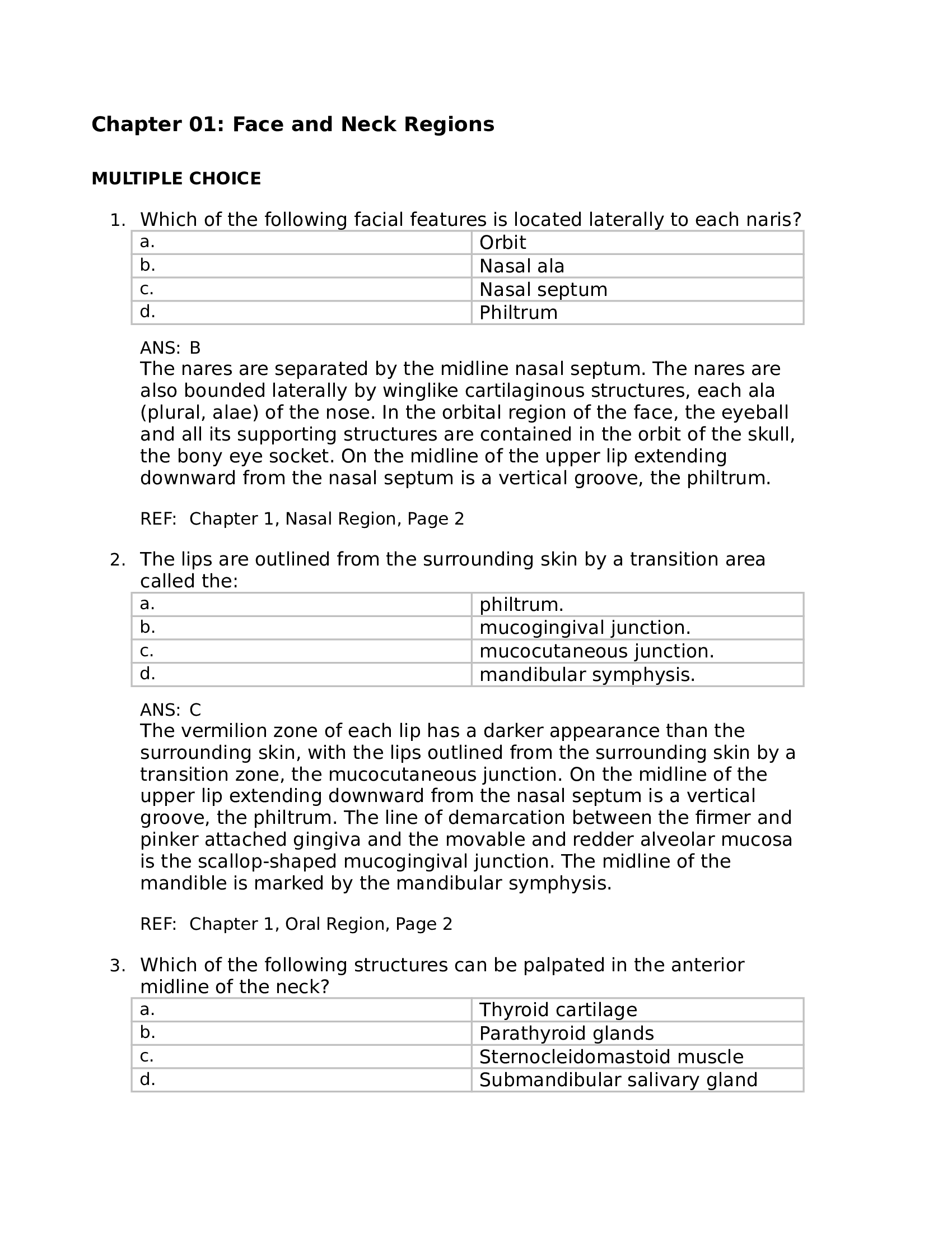
Chapter 01: Face and Neck Regions
MULTIPLE CHOICE
1. Which of the following facial features is located laterally to each naris?
a.
Orbit
b.
Nasal ala
c.
Nasal septum
d.
Philtrum
ANS: B
The nares are separated by the midline nasal septum. The nares are
also bounded laterally by winglike cartilaginous structures, each ala
(plural, alae) of the nose. In the orbital region of the face, the eyeball
and all its supporting structures are contained in the orbit of the skull,
the bony eye socket. On the midline of the upper lip extending
downward from the nasal septum is a vertical groove, the philtrum.
REF: Chapter 1, Nasal Region, Page 2
2. The lips are outlined from the surrounding skin by a transition area
called the:
a.
philtrum.
b.
mucogingival junction.
c.
mucocutaneous junction.
d.
mandibular symphysis.
ANS: C
The vermilion zone of each lip has a darker appearance than the
surrounding skin, with the lips outlined from the surrounding skin by a
transition zone, the mucocutaneous junction. On the midline of the
upper lip extending downward from the nasal septum is a vertical
groove, the philtrum. The line of demarcation between the firmer and
pinker attached gingiva and the movable and redder alveolar mucosa
is the scallop-shaped mucogingival junction. The midline of the
mandible is marked by the mandibular symphysis.
REF: Chapter 1, Oral Region, Page 2
3. Which of the following structures can be palpated in the anterior
midline of the neck?
a.
Thyroid cartilage
b.
Parathyroid glands
c.
Sternocleidomastoid muscle
d.
Submandibular salivary gland
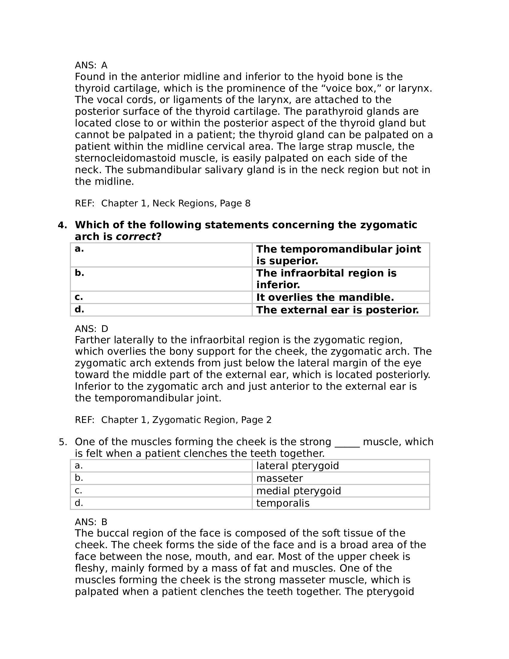
ANS: A
Found in the anterior midline and inferior to the hyoid bone is the
thyroid cartilage, which is the prominence of the “voice box,” or larynx.
The vocal cords, or ligaments of the larynx, are attached to the
posterior surface of the thyroid cartilage. The parathyroid glands are
located close to or within the posterior aspect of the thyroid gland but
cannot be palpated in a patient; the thyroid gland can be palpated on a
patient within the midline cervical area. The large strap muscle, the
sternocleidomastoid muscle, is easily palpated on each side of the
neck. The submandibular salivary gland is in the neck region but not in
the midline.
REF: Chapter 1, Neck Regions, Page 8
4. Which of the following statements concerning the zygomatic
arch is correct?
a.
The temporomandibular joint
is superior.
b.
The infraorbital region is
inferior.
c.
It overlies the mandible.
d.
The external ear is posterior.
ANS: D
Farther laterally to the infraorbital region is the zygomatic region,
which overlies the bony support for the cheek, the zygomatic arch. The
zygomatic arch extends from just below the lateral margin of the eye
toward the middle part of the external ear, which is located posteriorly.
Inferior to the zygomatic arch and just anterior to the external ear is
the temporomandibular joint.
REF: Chapter 1, Zygomatic Region, Page 2
5. One of the muscles forming the cheek is the strong _____ muscle, which
is felt when a patient clenches the teeth together.
a.
lateral pterygoid
b.
masseter
c.
medial pterygoid
d.
temporalis
ANS: B
The buccal region of the face is composed of the soft tissue of the
cheek. The cheek forms the side of the face and is a broad area of the
face between the nose, mouth, and ear. Most of the upper cheek is
fleshy, mainly formed by a mass of fat and muscles. One of the
muscles forming the cheek is the strong masseter muscle, which is
palpated when a patient clenches the teeth together. The pterygoid
muscles are also located on each side of the face near the mouth, and
the temporalis is located in the temporal region.
REF: Chapter 1, Buccal Region, Page 2
6. Just inferior to the zygomatic arch and just anterior to the external ear
is the:
a.
temporomandibular joint.
b.
infraorbital region.
c.
mental region.
d.
parotid gland.
ANS: A
Inferior to the zygomatic arch and just anterior to the external ear is
the temporomandibular joint. The infraorbital region of the face is
located inferior to the orbital region and lateral to the nasal region.
Farther laterally is the zygomatic region, which overlies the bony
support for the cheek, the zygomatic arch. The zygomatic arch extends
from just below the lateral margin of the eye toward the middle part of
the external ear. The chin is the major feature of the mental region of
the face; the bone underlying the mental region is the mandible, or
lower jaw. The parotid is located irregularly from the zygomatic arch
down to the posterior border of the lower jaw.
REF: Chapter 1, Zygomatic Region, Page 2
7. The _____ salivary gland is located irregularly from the zygomatic arch
down to the posterior border of the lower jaw.
a.
parotid
b.
submandibular
c.
von Ebner
d.
sublingual
ANS: A
The parotid is located irregularly from the zygomatic arch down to the
posterior border of the lower jaw. Both the submandibular salivary and
sublingual are in the neck region. The von Ebner refers to minor glands
located deep to the circumvallate lingual papillae on the dorsal surface
of the tongue.
REF: Chapter 1, Buccal Region, Page 2
8. The _____ extends from just below the lateral margin of the eye toward
the middle part of the external ear.
a.
temporomandibular joint
b.
zygomatic arch
c.
labial commissure
d.
infraorbital region
ANS: B
The zygomatic arch extends from just below the lateral margin of the
eye toward the middle part of the external ear. Inferior to the
zygomatic arch and just anterior to the external ear is the
temporomandibular joint. The upper and lower lips meet at each corner
of the mouth at the labial commissure. The infraorbital region of the
face is located inferior to the orbital region and lateral to the nasal
region.
REF: Chapter 1, Zygomatic Region, Page 2
9. The main feature of the _____ region of the face is the external nose.
a.
infraorbital
b.
zygomatic
c.
nasal
d.
temporal
ANS: C
The main feature of the nasal region of the face is the external nose.
The infraorbital region of the face is located inferior to the orbital
region and lateral to the nasal region. Farther laterally is the zygomatic
region, which overlies the bony support for the cheek, the zygomatic
arch. The temporal region is on the lateral side of the head at the
temple.
REF: Chapter 1, Nasal Region, Page 1
10. Which of the following orofacial structures is located in the midline of
the face or neck?
a.
Philtrum
b.
Submandibular salivary gland
c.
Naris and ala
d.
Parotid salivary gland
ANS: A
On the midline of the upper lip extending downward from the nasal
septum is a vertical groove, the philtrum. The submandibular can also
be palpated in a patient in the neck region but not in the midline.
Inferior to the apex on each side of the nose is a nostril, or naris
(plural, nares). The nares are also bounded laterally by winglike
cartilaginous structures, each ala (plural, alae) of the nose. The parotid
is located irregularly from the zygomatic arch down to the posterior
border of the lower jaw and not in the midline.
REF: Chapter 1, Oral Region, Page 2
11. The _____ region includes the forehead and area above the eyes.
a.
infraorbital
b.
orbital
c.
temporal
d.
frontal
ANS: D
The frontal region of the face includes the forehead and the area above
the eyes. The infraorbital region of the face is located inferior to the
orbital region and lateral to the nasal region. In the orbital region of the
face, the eyeball and all its supporting structures fare contained in the
orbit of the skull, the bony eye socket. The temporal region is on the
lateral side of the head at the temple.
REF: Chapter 1, Frontal Region, Page 1
12. Which of the following may be involved in the disruption of the
vermilion zone and may make it hard to determine the exact location
of its mucocutaneous junction between the lips and the surrounding
skin?
a.
Scar tissue
b.
Hypercalcification
c.
Cleft palate
d.
Symmetry
ANS: A
Disruption of the vermilion zone may make it hard to determine the
exact location of its mucocutaneous junction between the lips and the
surrounding skin. These changes may be due to scar tissue from past
traumatic incidents, developmental disturbances, or cellular changes in
the tissue such as occur with solar damage. These changes may also
composed of cementum. The bonelike cementum is the part of the
tooth that attaches to the periodontal ligament.
REF: Chapter 2, Jaws, Alveolar Processes, and Teeth, Page 12
14. Just distal to the last tooth of the maxillary arch is a tissue-covered
elevation of the bone called the:
a.
maxillary arch.
b.
maxillary tuberosity.
c.
canine eminence.
d.
retromolar pad.
ANS: B
Just distal to the last tooth of the maxillary arch is a tissue-covered
elevation of the bone, the maxillary tuberosity. Similarly, on the lower
jaw is a dense pad of tissue located just distal to the last tooth of the
mandibular arch, the retromolar pad. The alveolar processes with the
teeth in the alveoli are also called dental arches, the maxillary arch
and mandibular arch. The facial part of the alveolus of each canine, the
vertically placed canine eminence, is especially prominent on each side
of the maxilla.
REF: Chapter 2, Dental Arches, Page 12
15. On which of the following orofacial tissue is the linea alba located?
a.
Attached gingiva
b.
Marginal gingiva
c.
Labial mucosa
d.
Buccal mucosa
ANS: D
A variation that can be noted on the buccal mucosa is the linea alba.
This is a white ridge of hyperkeratinization that extends horizontally at
the level where the maxillary and mandibular teeth come together and
occlude; similar ridges of white tissue can sometimes be present on
the tongue perimeter.
REF: Chapter 2, Clinical Considerations with Oral Mucosa, Page 10
16. The structures closest to the lips are termed _____ or labial.
a.
lingual
b.
facial
c.
buccal
d.
palatal
ANS: B
The structures closest to the facial surface are facial. The facial
structures closest to the lips are labial. The facial structures close to
the inner cheek are buccal. The structures closest to the tongue are
lingual. The lingual structures closest to the palate are palatal.
REF: Chapter 2, Oral Cavity Divisions, Page 9
17. The pink labial mucosa or buccal mucosa meets the redder _____ at the
mucobuccal fold.
a.
marginal gingiva
b.
attached gingiva
c.
alveolar mucosa
d.
interdental papilla
ANS: C
Deep within each vestibule is the vestibular fornix, where the pink
labial mucosa or buccal mucosa meets the redder alveolar mucosa at
the mucobuccal fold. The gingival tissue that tightly adheres to the
alveolar process surrounding the roots of the teeth is the attached
gingiva. At the gingival margin of each tooth is the marginal gingiva,
which forms a cuff above the neck of the tooth. The interdental gingiva
is the gingival tissue between adjacent teeth adjoining attached
gingiva, with each individual extension being an interdental papilla.
REF: Chapter 2, Oral Vestibules, Page 10
18. The _____ is a white ridge of raised callused tissue that extends
horizontally at the level where the maxillary and mandibular teeth
come together and occlude.
a.
linea alba
b.
buccal fat pad
c.
parotid papilla
d.
labial frenum
ANS: A
The linea alba is a white ridge of hyperkeratinization that extends
horizontally at the level where the maxillary and mandibular teeth
come together and occlude; similar ridges of white tissue can
sometimes be present on the tongue perimeter. The buccal mucosa
covers a dense pad of underlying fat tissue at the posterior part of
each vestibule, the buccal fat pad. On the inner part of the buccal
mucosa, just opposite the maxillary second molar, is a small elevation
of tissue is the parotid papilla. The labial frenum (plural, frena) is a fold
of tissue located at the midline between the labial mucosa and the
alveolar mucosa on the upper and lower dental arches.
REF: Chapter 2, Clinical Considerations with Oral Mucosa Features, Page 10
19. Each body of the maxilla is superior to the teeth and contains the _____
sinuses.
a.
sphenoidal
b.
ethmoidal
c.
frontal
d.
maxillary
ANS: D
Each body of the maxilla, a facial bone, is superior to the teeth and
contains the maxillary sinus. The other sinuses listed—the sphenoidal,
ethmoidal, and frontal—are contained within the cranial bones.
REF: Chapter 2, Jaws, Alveolar Processes, and Teeth, Page 10
20. The facial part of the alveolus of the ____, the vertically placed
eminence, is especially prominent on the maxilla.
a.
premolar
b.
canine
c.
molar
d.
incisor
ANS: B
The facial part of the alveolus of each canine, the vertically placed
canine eminence, is especially prominent on each side of the maxilla.
The alveolar process, or alveolar bone, is the bony extension for both
the maxilla and mandible that contains each tooth socket of the teeth
or alveolus (plural, alveoli).
REF: Chapter 2, Jaws, Alveolar Processes, and Teeth, Page 10
21. Surrounding the teeth in the alveoli and covering the alveolar
processes is the _____, which is composed of a firm pink tissue.
a.
gingiva
b.
minor salivary glands
c.
Fordyce spots
d.
linea alba
ANS: A
Surrounding the maxillary and mandibular teeth in the alveoli and
covering the alveolar processes are the soft tissue gums, or gingiva (or
more accurately, but not commonly, by the dental community,
gingivae), composed of a firm pink mucosa. Minor salivary glands are
deep within the oral mucosa throughout the oral cavity. Fordyce spots
are visible as small, yellowish elevations on the oral mucosa; they
represent deeper deposits of sebum from trapped or misplaced
sebaceous gland tissue, usually associated with hair follicles. The linea
alba is a white ridge of hyperkeratinization that extends horizontally at
the level where the maxillary and mandibular teeth come together and
occlude; similar ridges of white tissue can sometimes be present on
the tongue perimeter.
REF: Chapter 2, Gingival Tissue, Page 13
22. What is the midline ridge of tissue on the hard palate?
a.
Incisive papilla
b.
Palatine rugae
c.
Median palatine raphe
d.
Uvula
ANS: C
The median palatine raphe is a midline ridge of tissue on the hard
palate. The incisive papilla is a small bulge of tissue at the most
anterior part of the hard palate. The palatine rugae are firm, irregular
ridges of tissue on the hard palate. A midline muscular structure, the
uvula of the palate, hangs down from the posterior margin of the soft
palate.
REF: Chapter 2, Palate, Page 15
23. What are the small, elevated structures of specialized mucosa located
on the dorsal surfaces of the tongue, some of which are associated
with taste buds?
a.
Lingual papilla
b.
Sulcus terminalis
c.
Parotid papilla
d.
Foramen cecum
ANS: A
The dorsal surface of the tongue has small, elevated structures of
specialized mucosa, the lingual papillae, some of which are associated
with taste buds. Posteriorly on the dorsal surface of the tongue is an
inverted V-shaped groove, the sulcus terminalis. Where the sulcus
terminalis points backward toward the throat is a small, pitlike
depression, the foramen cecum. On the inner part of the buccal
mucosa, just opposite the maxillary second molar, is a small elevation
of tissue is the parotid papilla.
REF: Chapter 2, Tongue, Page 15
24. What is the small bulge of tissue at the most anterior part of the hard
palate?
a.
Incisive papilla
b.
Palatine rugae
c.
Median palatine raphe
d.
Uvula
ANS: A
The incisive papilla is a small bulge of tissue at the most anterior part
of the hard palate. The palatine rugae are firm, irregular ridges of
Please Sign In to contact this author.